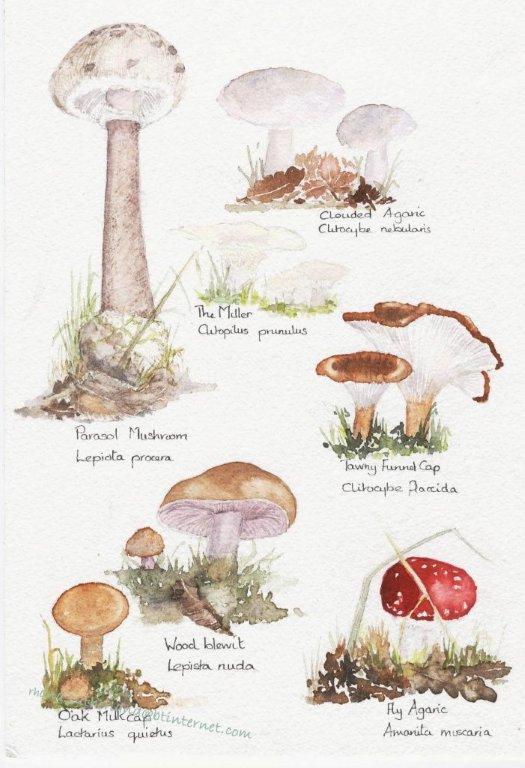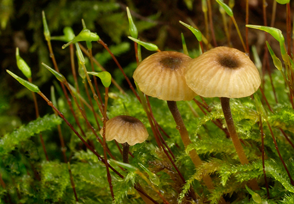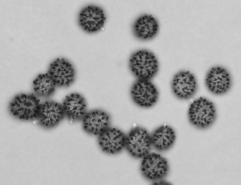
Russula ochroleuca spores x 1000
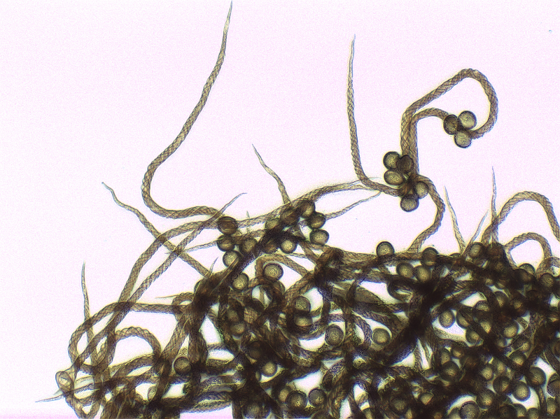
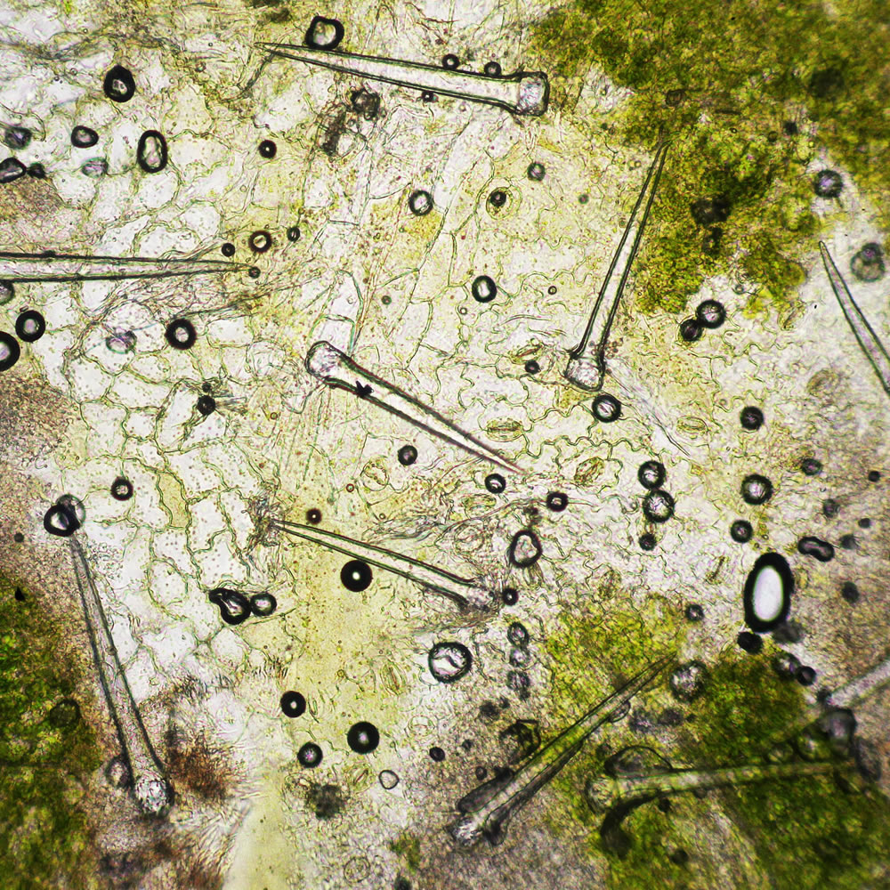
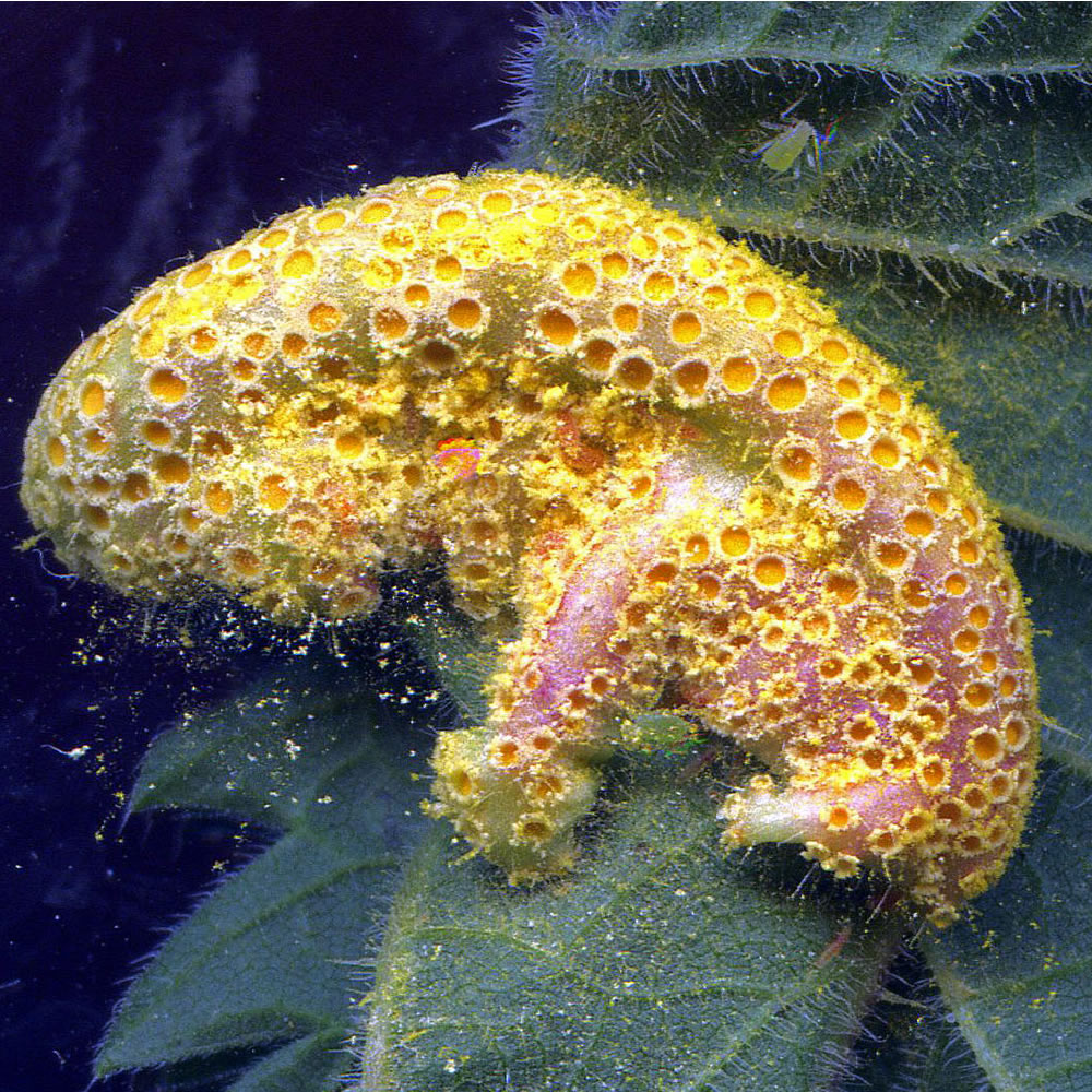
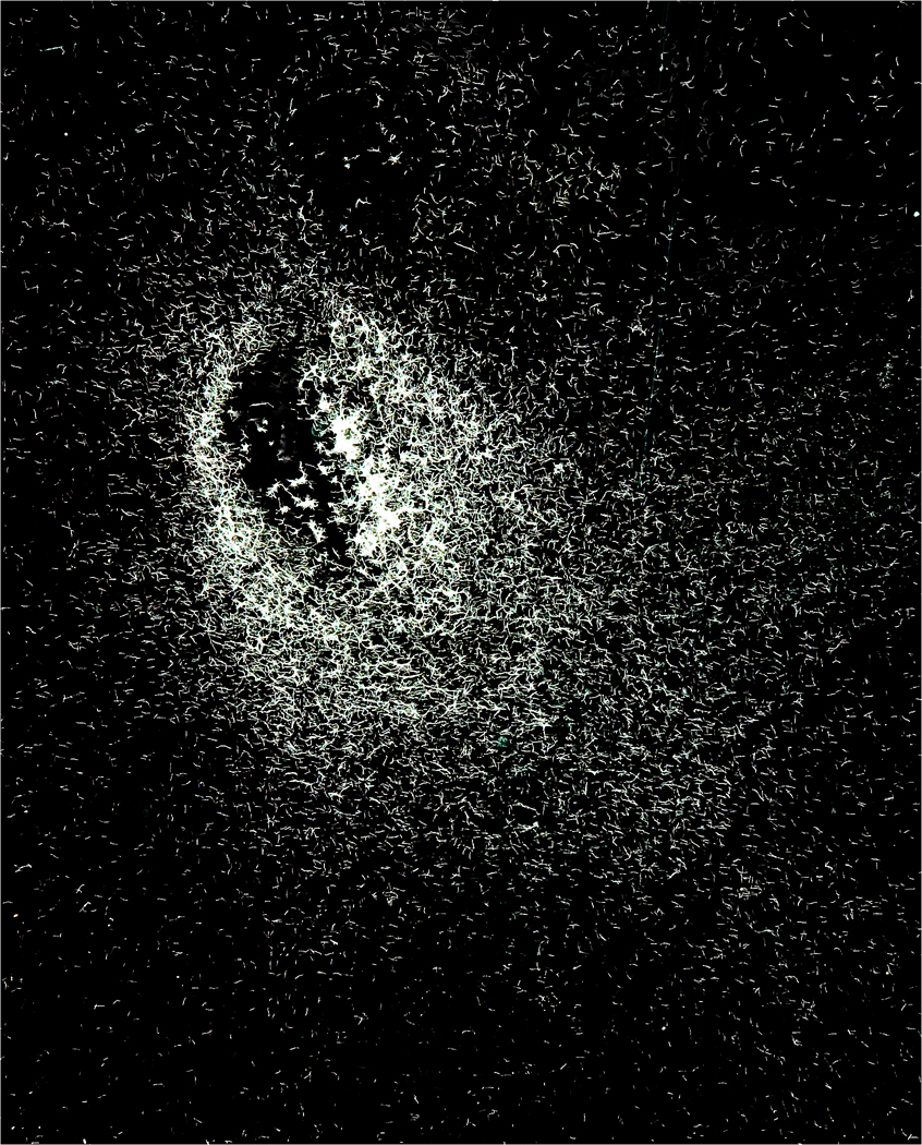
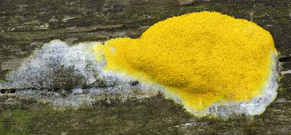
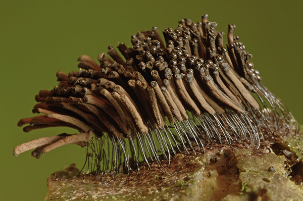
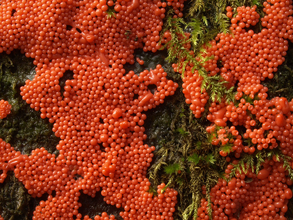
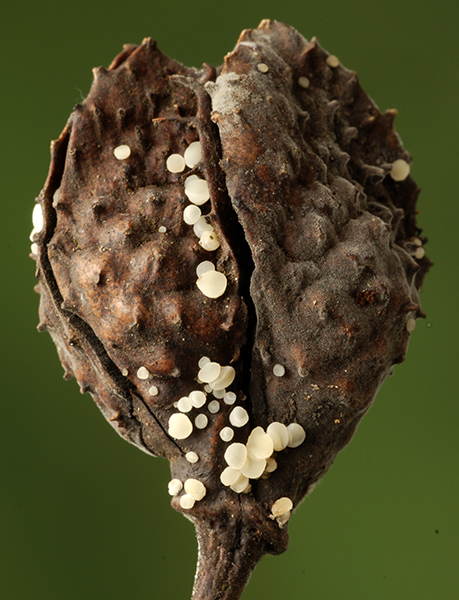
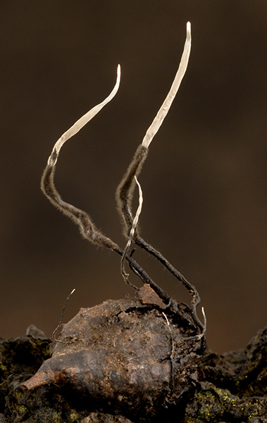
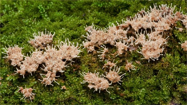
Helical structure of the threads (capillitium) of the Slime mould Metatrichia floriformis. The rapidly unwinding threads help to disperse the spores into the air. x400
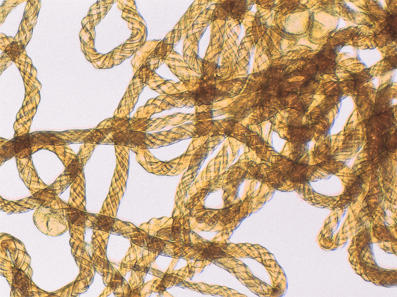
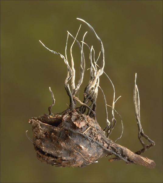
Helical capillitium of Metatrichia floriformis x 1000
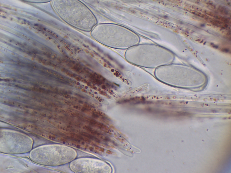
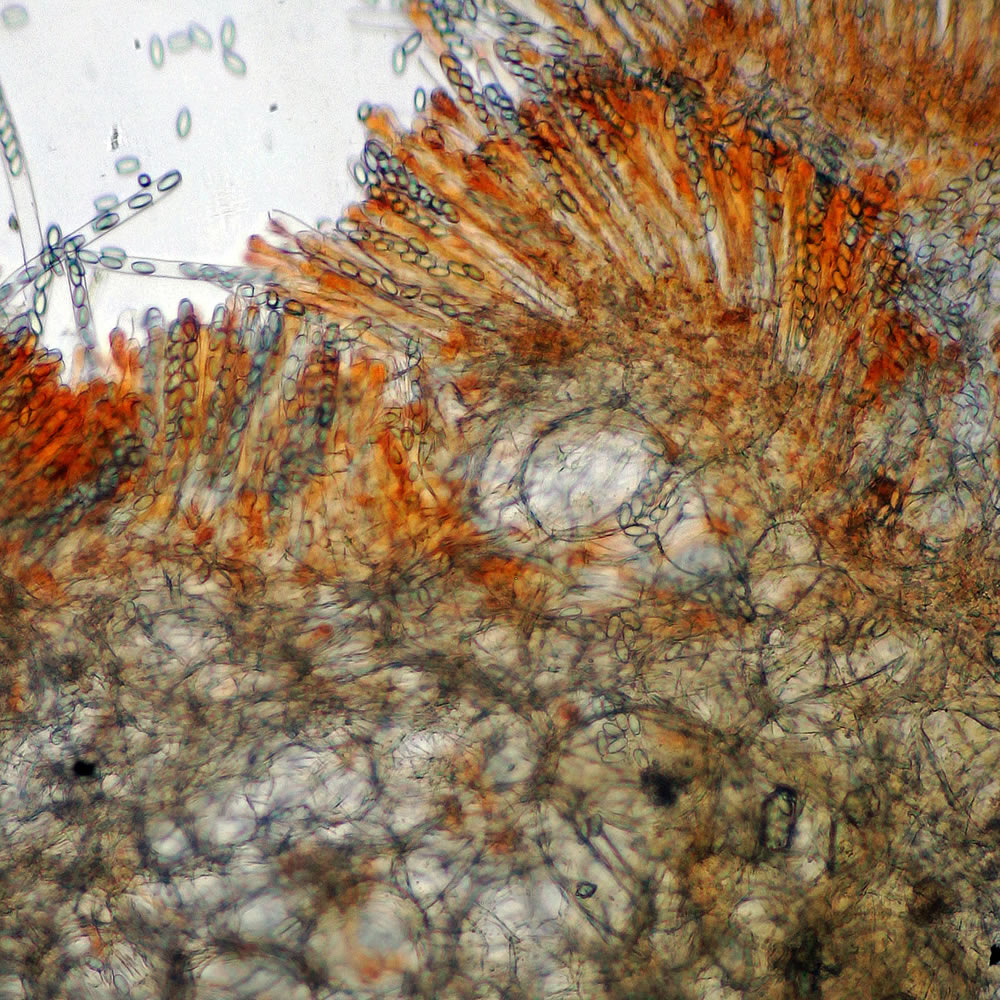
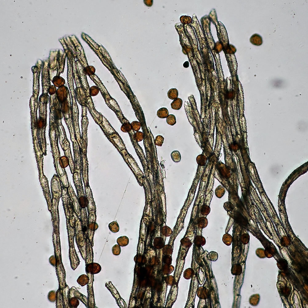
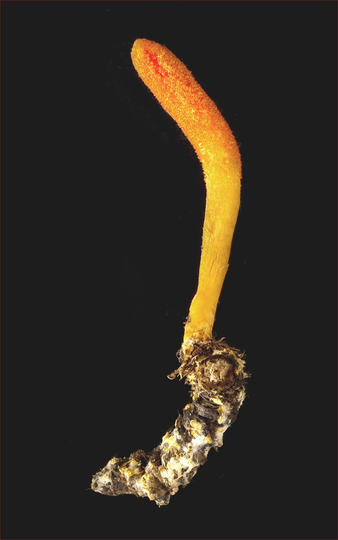
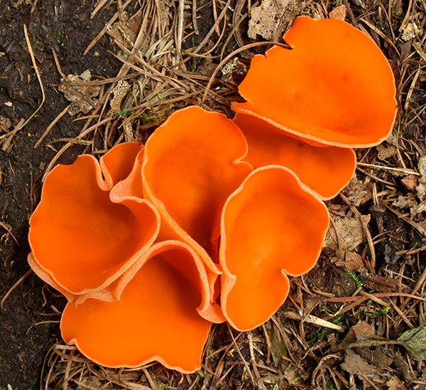
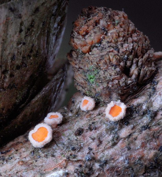
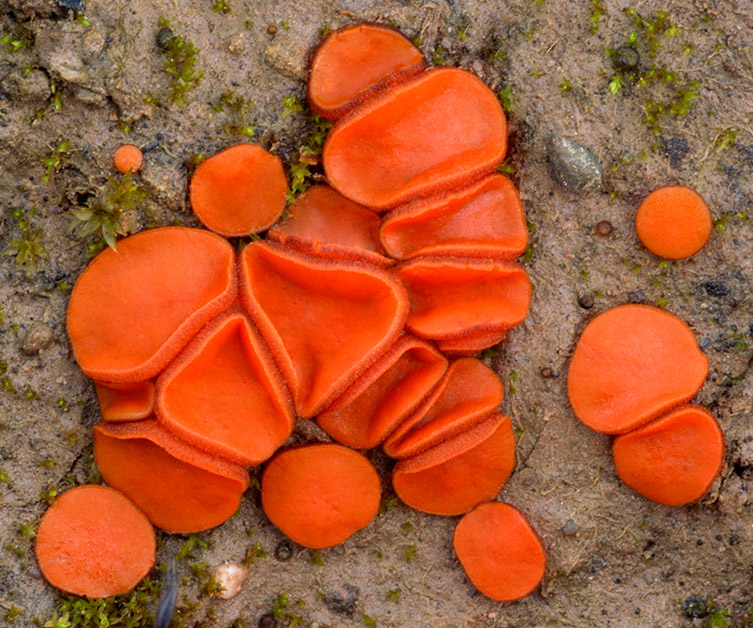
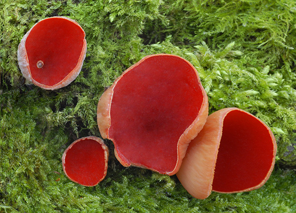
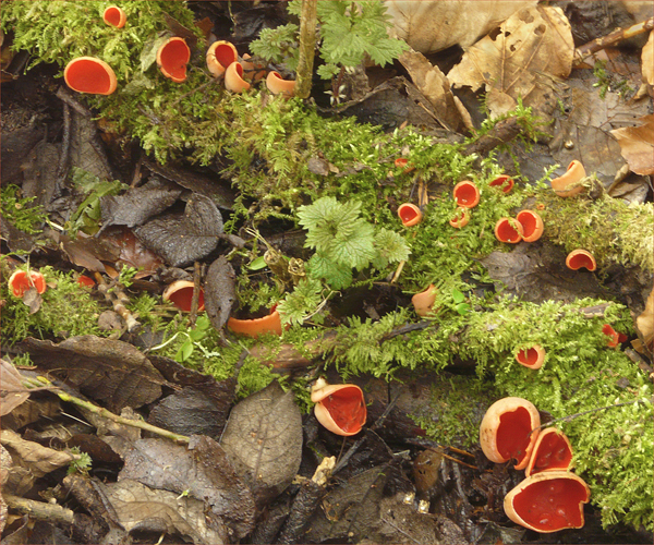
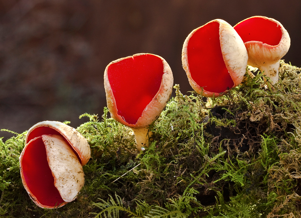
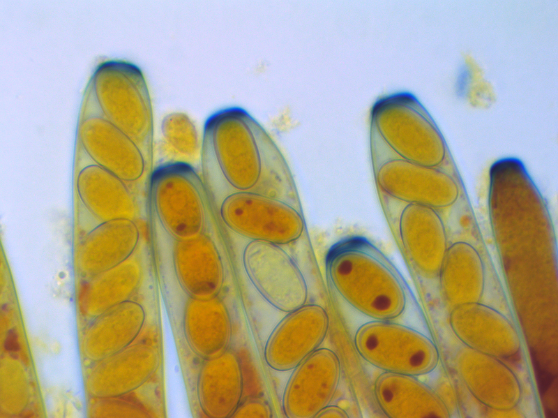
Sarcocypha austrica spores x 1000. Red carotenoid pigment can be seen in the paraphyses
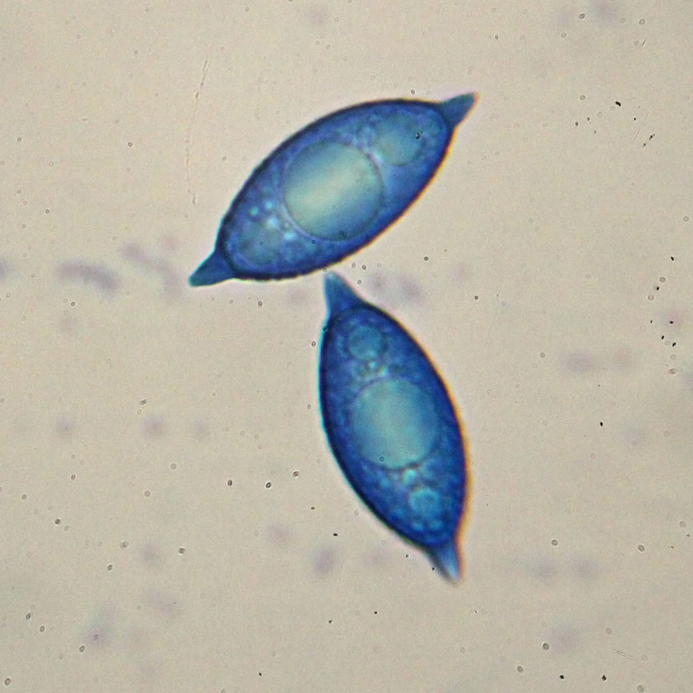
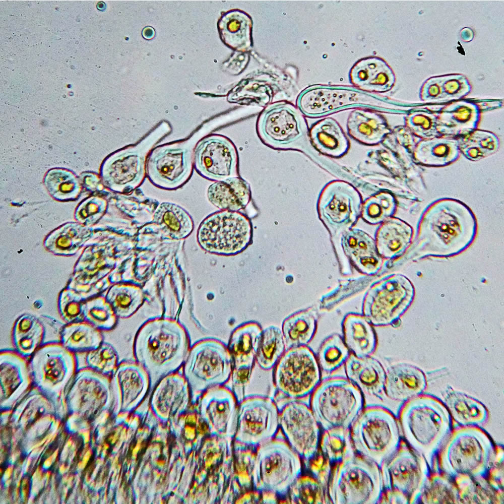
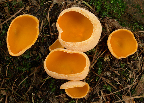
Peziza sp. (Baral’s Iodine) x 1000
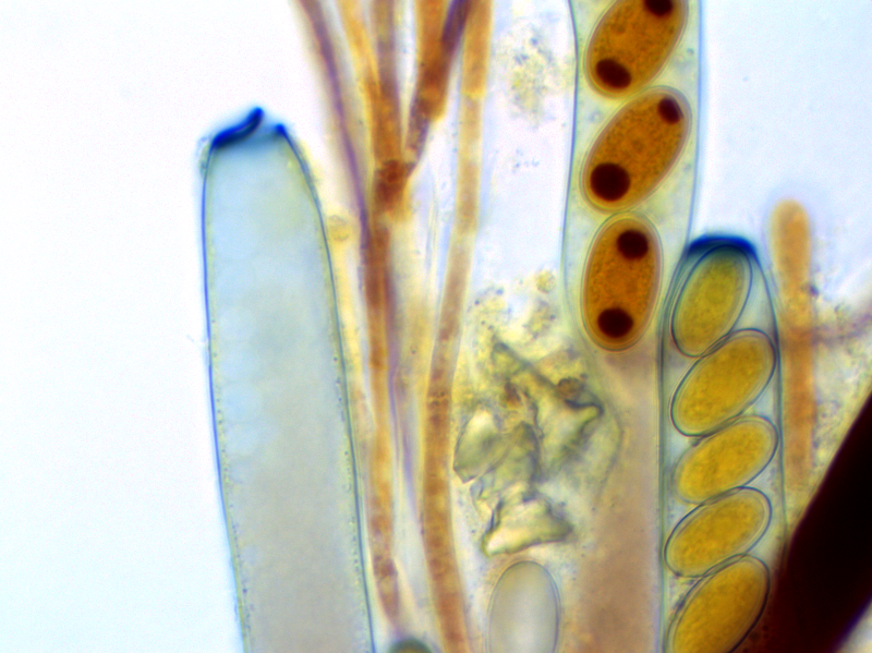
Peziza sp asci stained with Baral’s Iodine x 1000. The empty ascus on the left has released it’s spores and the blue lid remains open.